Michelle’s brain tumour story

In mid-2021, Michelle (54) from Princes Risborough, Buckinghamshire, found herself unexpectedly struggling with her vision.
Having lived with multiple sclerosis (MS) for over 23 years, Michelle originally put her eyesight struggles down to being a new symptom of her condition, or a possible MS reaction to ankle fusion surgery she had undergone to treat her arthritis.
Initially not thinking much of it, Michelle decided to visit the optician after her vision worsened over the course of a few months.
After some routine tests, the optician speculated that glaucoma – a common eye condition where the optic nerve becomes damaged – could be the cause of her loss of peripheral vision.
The diagnosis came as a complete shock
Michelle was referred to a consultant ophthalmologist for a series of tests to confirm whether or not her problems were in fact caused by glaucoma. During this process, it was recommended she have a brain CT scan, a routine measure intended simply to rule out any further issues.
However, one week later, she was called in to see her consultant who told her that the scans had shown a 3 ½ cm tumour on the brain.
Having had no other symptoms that are commonly associated with brain tumours, such as headaches or nausea, this revelation came as a complete shock to Michelle.
Recalling the experience, Michelle said: “Receiving the news that it was a brain tumour causing my vision issues left me in total shock. I had never expected it to be something so serious. But, having lived with MS and arthritis for a number of years already, I was determined that this would be another challenge to try and overcome.”
State-of-the-art technology
In March of this year, Michelle was referred to Mr Ian Sabin, Consultant Neurosurgeon at The Wellington Hospital, part of HCA Healthcare UK.
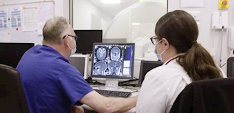
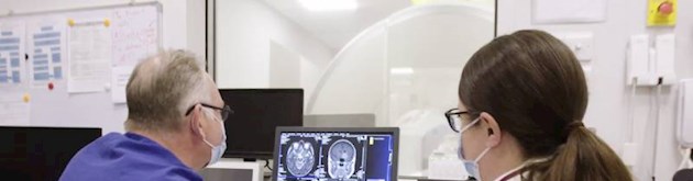
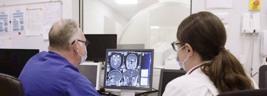
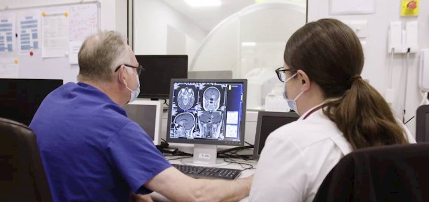
A series of consultations followed, assessing the best treatment options for removing the tumour. The scans were showing the tumour to be wrapped around the carotid artery as well as the optic nerve – which was causing the deterioration of her vision. Removal was going to be a complex procedure, but one that would be made more accessible using state-of-the-art iMRI technology. This would enable live visualisation and assessment of changes to the brain during surgery.
Having surgery with the iMRI
Excited at the prospect of having access to the iMRI scanner at The Wellington Hospital, Michelle decided to proceed with the treatment and on 30th May 2022, Michelle underwent surgery to remove the tumour – the first patient to benefit from this technology at HCA UK.
The operation went smoothly, and Michelle was kept in intensive care for 4 days. After making good progress with her speech and cognitive function (which can be temporarily affected by brain surgery), she was moved onto a ward for a day before being sent home to make a full recovery.
At her six-week follow up appointment with Mr Sabin, he had only positive news to share.
Talking her through the scans from before, during and after the surgery, it was revealed that the outcome of the surgery had been extremely positive. The whole tumour had been successfully removed.
In a biopsy, the tumour was found to be benign, meaning that no further treatment would be required, and the chances of recurrence are very slim.
Michelle recalled: “I was beyond impressed with my experience at The Wellington Hospital. From day one, Mr Sabin gave me the honest advice I desperately wanted, reassuring me that I was in safe hands and making the right decision to opt for the iMRI surgery. “I am now happy to be back at home with my husband, and am steadily improving every day, which is all I could ever ask for.”
A long-term definitive treatment
Looking back on Michelle’s treatment, Mr Ian Sabin, Consultant Neurosurgeon at The Wellington Hospital, said:
“It was a pleasure to be able to offer Michelle the chance to be the first patient treated using the new iMRI technology at The Wellington Hospital.
While her surgery was certainly a complex procedure, with the tumour wrapped around the carotid artery and optic nerve, with the help of the iMRI technology, we were able to remove it in its entirety.
I’m pleased to say that Michelle’s vision, speech, and cognitive function has made a brilliant recovery. The tumour was found to be benign, and recurrence is small, so I am confident that the surgery will provide a long-term definitive treatment for her. We will of course monitor this with an annual MRI for the next five years.
I wish Michelle every success and a happy, healthy future.”
What is an iMRI?
The iMRI technology, provides high-definition images of the brain during surgery, helping to guide the surgery, distinguishing abnormal tissue from normal tissue and protecting critical structures, all within the operating room. The technology gives the surgeon confidence that they have achieved their treatment goal and reduces the need for the patient to have additional surgery in the early post-operative period.
A series of consultations followed, assessing the best treatment options for removing the tumour. The scans were showing the tumour to be wrapped around the carotid artery as well as the optic nerve – which was causing the deterioration of her vision. Removal was going to be a complex procedure, but one that would be made more accessible using state-of-the-art iMRI technology. This would enable live visualisation and assessment of changes to the brain during surgery.